
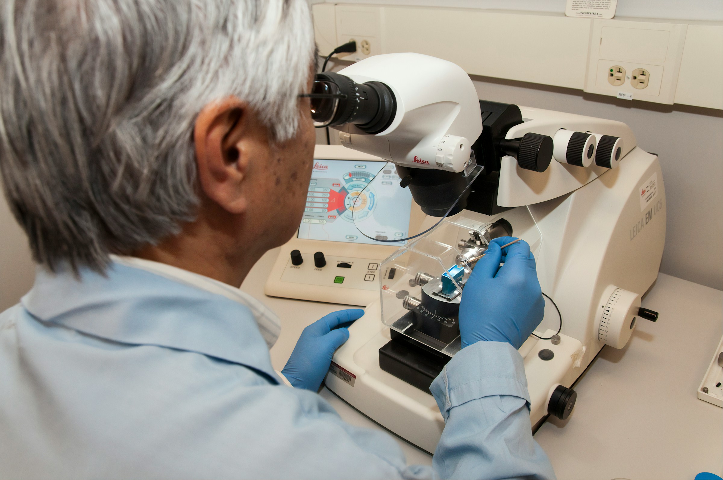
The image depicts a lab scientist with gray hair and blue gloves working with a Leica EM UC6 ultra-microtome microscope. The researcher is preparing thin sections of tissue, likely for microscopic analysis, a crucial process in cancer research and chemotherapy planning—especially for rare cancers like mesothelioma. The lab environment and digital interface highlight the role of technology in precision oncology.